Ever wonder how doctors help a racing heart slow down and find a safe rhythm? They use a set of guidelines called ACLS (Advanced Cardiac Life Support), which is like a step-by-step plan. First, they check breathing, pulse, and other important signs, kind of like double-checking that every door is locked before you head out. This careful process helps them spot tricky heart patterns. Then, they decide whether a simple maneuver or a careful electric reset is needed to steady the heart quickly.
Today, we’ll walk you through these trusted steps that show how clear, quick actions help save lives.
SVT Treatment in ACLS: Adult Tachycardia With a Pulse Algorithm
We start with a quick primary assessment where we check the Airway, Breathing, Circulation, Disability, and Exposure. Think of this first step like ensuring all the doors and windows in your home are locked before you set your alarm, it’s the basic safety check that makes everything else work smoothly.
After this essential review, we move on to the secondary assessment. Now, the focus shifts to spotting any reversible causes, often called H’s and T’s, gathering a clear medical history, and doing a targeted physical exam. This helps us figure out what might be causing the irregular heartbeat.
When the heart rate climbs above 150 beats per minute, we activate the Adult Tachycardia With a Pulse Algorithm. In cases of SVT, you usually see a narrow QRS complex (less than 0.12 seconds), although sometimes a wide QRS can appear. Recognizing these patterns on the ECG is key to making a correct diagnosis and planning the right treatment.
Once the ECG clues are clear, the algorithm guides the next steps. Doctors assess whether the patient is stable and then decide on the best move, be that trying out vagal maneuvers first, using medications, or even considering cardioversion. Simple reminders, like “if the rhythm is regular and the QRS is narrow, try vagal maneuvers,” help keep the process straightforward and safe.
This organized approach under ACLS guidelines ensures that every twist of an arrhythmia is handled carefully, protecting patients with quick and effective treatment every time.
Unstable SVT Management in ACLS: Cardioversion and Sedation
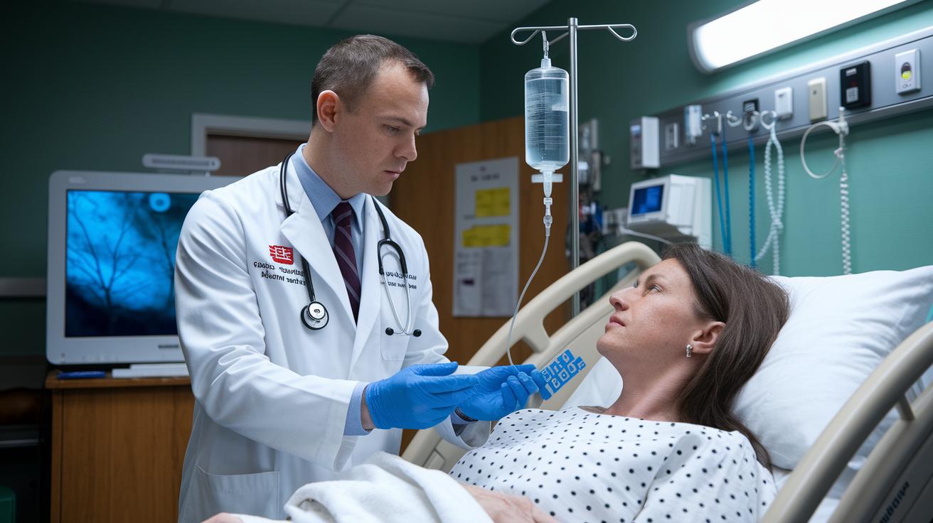
When someone shows signs of an unstable fast heartbeat, like low blood pressure, chest pain, confusion, or even shock, it’s time to act quickly. ACLS guidelines say that the next step is to perform synchronized cardioversion. That simply means giving a quick electrical shock while the patient is sedated. Think of it like rebooting a frozen computer to get everything working smoothly again.
If the heart beats at 150 beats per minute or more, or if the symptoms are very strong, this is the clear choice for treatment. But if a patient already has heart problems, even a slightly lower heartbeat can be worrisome. In those cases, doctors take extra care and monitor the patient closely while deciding on cardioversion.
Sedation plays a key role here. It helps calm the patient before the shock, much like taking a deep breath before an important moment. This careful, controlled approach makes a big difference in managing the emergency safely and effectively.
Stable SVT Treatment in ACLS: Vagal Maneuvers and Pharmacologic Conversion
When someone has stable SVT with a narrow QRS, the first thing to try is a simple vagal maneuver. This might be a Valsalva maneuver or a gentle carotid sinus massage. Think of it like giving your heart a little signal to take a deep breath and slow down.
If these maneuvers don’t fix the rhythm, the next step is to use medication. Adenosine is given quickly through an IV push. First, 6 mg is given along with a fast saline flush. If the heart is still racing, a second dose of 12 mg may be given. It’s like giving your heart a gentle nudge to get back on track.
For cases where the ECG shows a wide QRS with a monomorphic rhythm, similar treatments are used. In those situations, either adenosine or other IV medications such as procainamide, amiodarone, or sotalol might be used to safely convert the rhythm.
Every step in this process is designed to be both calm and effective, always keeping your heart’s safety first. Have you ever wondered how a simple maneuver might change everything?
SVT Treatment in ACLS: Adenosine Dosing Protocols

ACLS begins by giving a 6 mg IV shot through a vein close to the heart. Right after, a saline flush clears the line so the medicine can reach the heart quickly. Think of it like pressing a reset button for a lagging computer.
If the 6 mg dose doesn’t fix the abnormal heartbeat, a second shot of 12 mg is given. This extra step helps break the erratic rhythm when the initial dose isn’t enough.
It’s important to limit these rapid shots to just two doses. This simple plan explains both the method used and why we start with 6 mg, moving on to 12 mg only if needed.
| Dose | Description |
|---|---|
| 6 mg | Initial IV shot via a nearby vein with a quick saline flush |
| 12 mg | Second IV shot if the first dose doesn’t fix the heartbeat |
SVT Treatment in ACLS: ECG Criteria and Differential Diagnosis
Look for special ECG markers that help set SVT apart from other fast heart rhythms. Instead of only checking the QRS width, pay attention to the steady beat and the clear parts of the wave. In SVT, you usually see a consistent rhythm with easy-to-spot P waves, and generally, the signals from the upper and lower chambers work together normally.
Also, keep an eye out for hints like capture and fusion beats. These small clues can help tell SVT apart from ventricular tachycardia, especially when the usual ECG signs don't tell the whole story.
For example, imagine an ECG strip as a clear melody. A steady beat that plays like a smooth tune points to SVT, while a few off beats might suggest you need to take a closer look.
| ECG Marker | SVT Indicator |
|---|---|
| P wave visibility | Clear and steady P waves that come before each QRS complex |
| Capture beats | Sometimes a normal beat appears in the rapid rhythm |
| AV dissociation | Usually not seen in SVT |
SVT Treatment in ACLS: Secondary Assessment and H’s and T’s Evaluation

When performing an ACLS secondary assessment, providers focus on reversible causes, often called the H’s and T’s. They check for problems like low oxygen levels, potassium imbalances, decreased blood volume, blood clots, lung pressure issues, and toxins. It’s a bit like solving a mystery, small clues from a clear history, a targeted physical exam, and vital signs come together to point to the right diagnosis.
Imagine a patient with a fast heartbeat that might be caused by an unseen toxin. Start by asking a simple question during the medical history, like, “Have you been around any unusual substances lately?” This little inquiry can help guide the provider to think about toxic reasons behind the arrhythmia.
By following a clear set of steps for diagnosing and treating SVT during resuscitation, healthcare professionals can quickly spot the underlying issue. Gathering the right clinical details is as important as following a trusty map to arrive at the best treatment plan.
SVT Treatment in ACLS: Certification and Simulation Resources
ACLS, BLS, and PALS courses help you build skills in handling fast heartbeat events like SVT. These programs offer practice tests, realistic patient scenarios, and ECG rhythm challenges that boost your confidence during emergencies. Think of these exercises as training flights that prepare you to act quickly when a fast heartbeat happens.
Interactive training tools let you work through actual SVT cases and get feedback right away. For example, one simulation may ask if you should try a vagal maneuver or give adenosine. Did you know that in one practice session, doctors shortened their decision-making time by almost 20% by repeatedly practicing these scenarios? It really shows how hands-on training makes you more ready for the real thing.
Along with these practice tools, there are special discounts to help you learn even more. Right now, you can get 25% off with the code 4THJULY25 until 07/29/2025. You can also take advantage of group orders and get support from a dedicated help center. All of these resources work together to keep you updated on the most current ACLS-certified treatment methods, helping you feel safe and prepared when emergencies arise.
| Resource | Description |
|---|---|
| Simulation Scenarios | Interactive cases to build emergency response skills |
| Practice Tests | ECG rhythm challenges and case reviews |
Final Words
In the action, the article breaks down every step for tackling SVT using ACLS techniques. It covers everything from initial assessments and ECG clues to adenosine dosing, cardioversion, and even simulation training for practice.
Each section guides you through smart, secure methods for managing tachycardia, including how to handle challenging cases and use the right protocols. The insights provided ensure smooth svt treatment acls while keeping patient care the top priority. A clear path forward sparks confidence for improved health outcomes.
FAQ
Frequently Asked Questions
What does the SVT algorithm in ACLS involve?
The SVT algorithm in ACLS provides a step-by-step guide starting with a primary assessment, evaluating the ECG for rate and rhythm, then using vagal maneuvers for stable patients and cardioversion for unstable ones.
What is the adenosine dose for SVT treatment in ACLS?
The adenosine dose for SVT treatment starts with a 6 mg IV push followed by a saline flush; if needed, a 12 mg dose is given shortly after, according to ACLS protocols.
What is the treatment for stable SVT in ACLS?
The treatment for stable SVT in ACLS begins with vagal maneuvers like the Valsalva maneuver; if these fail, adenosine is administered to return the heart to normal rhythm.
How is unstable SVT treated in ACLS and what is the priority treatment?
Unstable SVT in ACLS is treated with immediate synchronized cardioversion along with appropriate sedation due to signs of hemodynamic instability such as low blood pressure or chest pain.
What medication is used for SVT in ACLS protocols?
In ACLS protocols, adenosine is the first-line medication used to convert SVT after vagal maneuvers, while other antiarrhythmics may be considered for cases unresponsive to initial treatment.
How is ventricular tachycardia with a pulse managed according to ACLS guidelines?
Management of VTach with a pulse in ACLS includes assessing hemodynamic stability and may involve the use of antiarrhythmic medications or synchronized cardioversion, following close monitoring of the patient’s condition.